Cat # changed from RD194349200R to RD194349220R
Type
Sandwich ELISA, Biotin-labelled antibody
Applications
Whole blood
Shipping
At ambient temperature. Upon receipt, store the product at the temperature recommended below.
Storage/Expiration
Store the complete kit at 2–8°C. Under these conditions, the kit is stable until the expiration date (see label on the box).
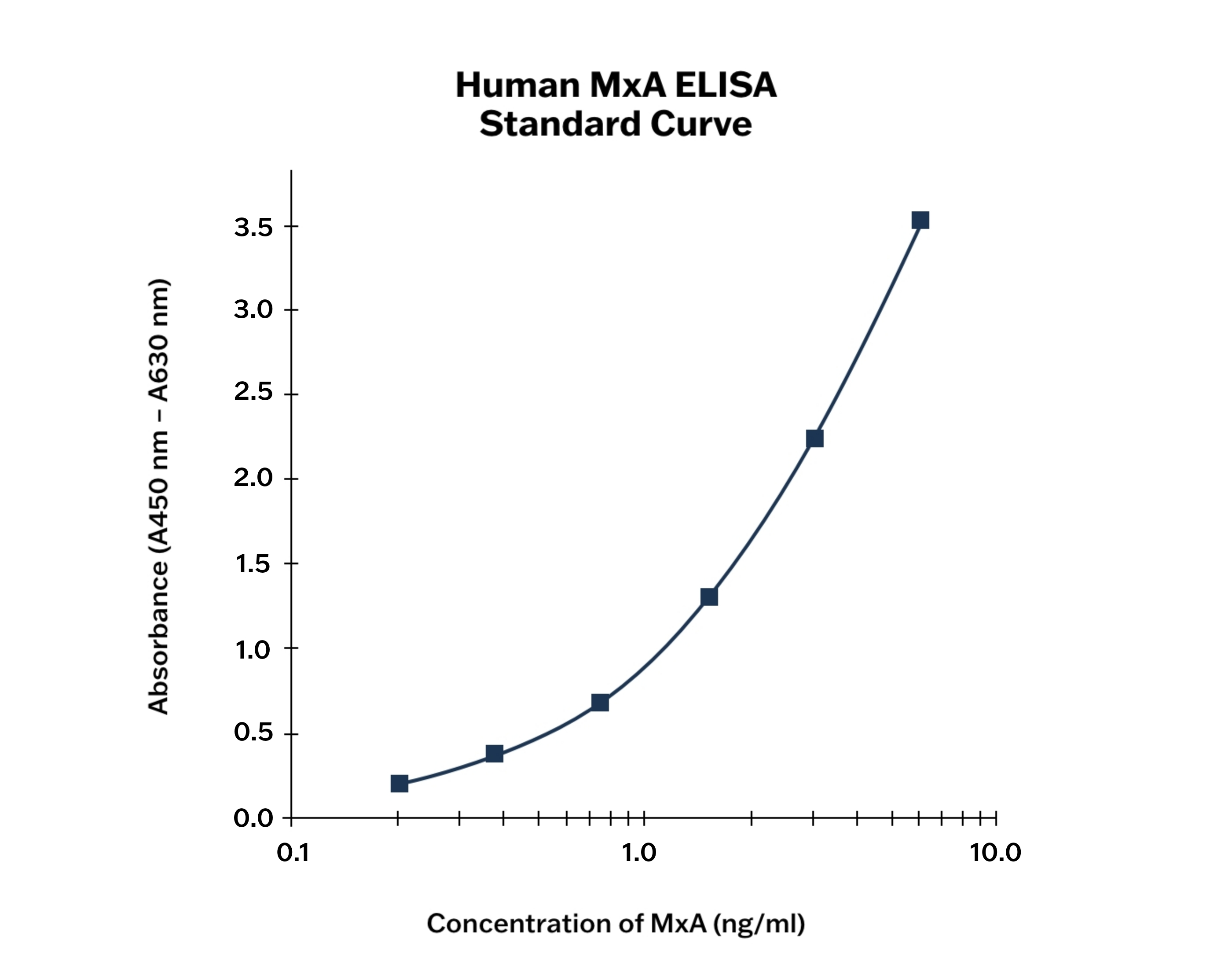
Calibration Curve
Calibration Range
0.188–6 ng/ml
Limit of Detection
0.001 ng/ml
Intra-assay (Within-Run)
n = 8; CV = 5.45%
Inter-assay (Run-to-Run)
n = 6; CV = 5.45%
Spiking Recovery
89.92%
Dilution Linearity
94.63%
Features
- It is intended for research use only.
- The total assay time is less than 3 hours.
- The kit measures MxA protein in whole blood (cell lysate).
- Assay format is 96 wells.
- Standard is native MxA protein based.
- Components of the kit are provided ready to use, concentrated or lyophilized
Research topic
Immune Response, Infection and Inflammation, Sepsis, COVID-19
Summary
Human MxA protein (Myxovirus resistance protein 1), encoded by the MX1 gene, is a 76-kDa protein composed of 662 amino acid residues and is a member of the dynamin superfamily of large GTPases. The MxA protein plays a crucial role in antiviral defense within cells, providing protection against a broad range of viruses, including influenza, parainfluenza, measles, coxsackie, hepatitis B, and Thogoto viruses.
The viruses are inhibited by MxA protein at an early stage in their life cycle, soon after host cell entry and before genome amplification. The human MxA protein is accumulated in the cytoplasm and endoplasmic reticulum. The membrane compartment of endoplasmatic reticulum seems to provide an interaction platform that facilitates viral target recognition. MxA appears to detect viral infection by sensing and trapping nucleocapsid structures, and becoming the viral components unavailable for the generation of new virus particles.The expression of viral MxA protein is induced exclusively and, in a dose-dependent manner, by IFN-alpha and IFN-beta, but not by IFN-gamma, IL-1, TNF-alpha or other cytokines.
MxA protein may offer advantages as a laboratory marker because of its very low basal concentration, increasing within 1-2 hours of infection and long half-life being approximately 2-3 days. In mononuclear cells stimulated with high doses of leukocyte IFN-alpha, MxA mRNA levels increased tenfold within 4 hours, and elevated MxA protein levels persists over the next 48 hours. MxA protein with its low basal concentration and long half-life, offers advantages as a marker for viral infection. Clinical studies have reported on MxA protein in peripheral blood mononuclear cells as a marker distinguishing viral from bacterial disease, and as a reliable marker for type I IFN bioavailability during IFN treatment in patients with multiple sclerosis (MS) according to the recommendation from EMA (European Medicine Agency).
Main Clinical Use of MxA
- Differentiating viral from bacterial infections
- Detection and assessment of active phase of Viral Infections
- Immune response assessment
- Autoimmune and inflammatory disease activity marker
- Therapeutic monitoring in interferon therapy
Find documents for the lot
Example Instructions for Use (RUO)
Example Instructions for Use (RUO)
Safety Information (RUO)
MSDS (RUO)
MSDS (RUO)
Product Brochure
MxA in managing infections and immune responses