United States set
language
Menu Shopping cart $0 Search

Raman Microscopy

BioRam® Photonic Fingerprinting

Amazingly Raman microscopy can indeed identify and analyze individual cells – alive or fixed – providing detailed information about the actual metabolic status of the cell.

4145

BioRam is a confocal microscope which combines Raman spectroscopy with laser technology to meet the specific requirements of the biological samples with broad spectrum of applications such as Cancer research, Stem Cell Research, Drug discovery, Quality control and last but not least Clinical practice.

Reinvent cell analysis

  • Label-free
  • Non-invasive
  • Specific & fast
  • Sample saving

Support healthiness

  • Cell-based therapeutics
  • Stem cell therapy
  • Regenerative medicine
  • Drug and compound safety
  • Tumor diagnosis

System introduction


About Raman Spectroscopy

About Raman Spectroscopy

Identifying cells from their biophotonic profile

Raman spectroscopy is based on the Raman effect: when a molecule is exposed to laser light, a small fraction is scattered with a shift in frequency compared to the incident light. This shift is highly specific for each molecule – as unique as a fingerprint.

4206
Raman spectra from airborne microorganism. Colonies were scraped off the Agar plate and resolved in buffer. Raman spectra were taken from individual living bacteria and fungi utilizing the simultaneous trapping effect of the Raman Laser configuration. Raman spectra of colored colonies differ at wavenumbers corresponding to carotenoids.

Raman spectroscopy allows to identify and analyze cells with a high degree of precision and specificity without the need of biochemical markers, fluorescent labels, antibodies or beads. It works within liquids under culture conditions and enables investigation of cellular components unaffected and preserved for further use.

Raman spectroscopy has long been used successfully as a contact-free technique for material characterization. More recently, scientists have demonstrated its applicability to the field of biology and medicine: several research teams have independently shown that the combined Raman spectra of all biopolymers within a cell form distinct clusters according to cell type and state of differentiation and activity.

Advantages & Applications

Advantages & Applications

4288

Advantages of BioRam® – Raman spectroscopy for everyone

With BioRam®, physicians, pharmacists and biologists gain uncomplicated access to the advantages of Raman spectroscopy. BioRam® is a powerful and effective tool for analyzing biological samples, including microorganisms, cells and tissue providing new insights into their nature and behaviour.

4306Safe and sound with BioRam®
Quality control & sample validation

  • Benefit from non-desctructive analysis of your product
  • Ensure the quality of cell based therapeutics
  • Guarantee cell viability and functionality
  • Screen for possible contamination
  • Increase safety for your patients

4307Innovative therapy is just a laser beam away
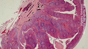
Tumor research & analysis

  • Discriminate tumor from non-tumor cells
  • Characterize tumor entities and staging
  • Discover tumor subpopulations
  • Screen for patient specific drugs

4308Shed light into cell behaviour
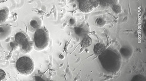
Cell culture & drug screening

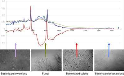
  • dentify different cell types and subpopulations
  • Monitor cell state and development
  • Screen for relevant biomolecules
  • Diagnose treatment and disease induced molecular changes

4309See the whole spectrum of cell development
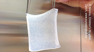
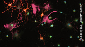
Cell cells & regenerative medicine

  • Characterize stem cell populations in-line
  • Detect and monitor cell differentiation
  • Prove functionality of differentiated cells
  • Depict cell composition in tissue products

4310
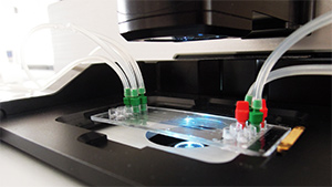
Benefits of BioRam® - Get into the flow

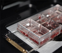
Integrated laser trapping properties arrest floating samples within the laser focus simultaneously with Raman spectra acquisition. Our unique combination of Raman microscopy, application-oriented software and modern microfluidics, supported by customized lab-on-a-chip systems, identifies, analyzes and sort cells in suspension in a simple, non-invasive way.

References

References

  • Buchacher T, Wiesinger-Mayr H, Vierlinger K, Ruger BM, Stanek G, Fischer MB, Weber V. Human blood monocytes support persistence, but not replication of the intracellular pathogen C. pneumoniae. BMC Immunol. 2014;15:60
  • Charwat V, Schutze K, Holnthoner W, Lavrentieva A, Gangnus R, Hofbauer P, Hoffmann C, Angres B, Kasper C. Potential and limitations of microscopy and Raman spectroscopy for live-cell analysis of 3D cell cultures. J Biotechnol. 2015 Jul 10;205:70–81
  • Smith R, Wright KL, Ashton L. Raman spectroscopy: an evolving technique for live cell studies. Analyst. 2016 Jun 21;141(12):3590-600
  • Steinke M, Gross R, Walles H, Gangnus R, Schutze K, Walles T. An engineered 3D human airway mucosa model based on an SIS scaffold. Biomaterials. 2014 Aug;35 (26):7355–62