United States set
language
Menu Shopping cart $0 Search

Tescan

We proudly introduce the Q-PHASE, a multimodal holographic microscope (MHM). With this instrument BioVendor and TESCAN expand into the field of advanced light microscopy. The Q-PHASE is a unique instrument for quantitative phase imaging (QPI) based on patented technology of Coherence-controlled holographic microscopy. [1,2]

4252
  • High quality quantitative phase imaging (QPI)
  • Full motorization
  • Multimodality (combination of holographic and fluorescence microscopy)
  • Label free and low phototoxicity
  • In-vitro cell monitoring
  • Useful in many biological and biotechnical applications (cell’s life cycles, cell motility or morphology changes, cell growth analysis and many others)


This product is only available in these countries: Austria, Germany, Switzerland

About

About

Coherence-controlled holographic microscopy

Raman spectroscopy is based on the Raman effect: when a molecule is exposed to laser light, a small fraction is scattered with a shift in frequency compared to the incident light. This shift is highly specific for each molecule – as unique as a fingerprint.

Principles of quantitative phase imaging

The time of propagation of light in a specific environment depends on the refractive index as well as the distance of the optical path. Therefore, when a light wave travels through a sample with varying refractive index and/or height, its wavefront is distorted and a change in the phase distribution of this wave occurs. The Q-PHASE is able to detect the phase distribution in the sample plane. This process of phase detection in a sample plane is usually referred to as quantitative phase imaging. Quantitative phase image can provide information on sample morphology, topography or cell dry-mass distribution. [5,6] Cell dry mass is quantified in pg/μm2 and can be calculated directly from phase values detected in each pixel. Quantitative phase imaging provides a very simple and sensitive way for monitoring of cell reactions to treatment and analyses of movement, growth, area, shape and many other parameters. Various color LUTs are often used for representation of phase images to easily distinguish different phase values.

6859

6860

Patented optical setup

The Q-PHASE microscope consists of two arms, object arm and reference arm. The arms have similar microscope setups with a common illumination system. The sample is placed into the object arm, and the so-called reference sample (blank) is placed into the reference arm. The beams in each arm pass through the inserted sample and are combined at the image plane of the microscope. Thanks to the Q-PHASE’s unique patented optical setup, the beams interfere and form a hologram even when illuminated with a halogen lamp or a LED. The hologram is then recorded by a detector and a quantitative phase image is extracted from the hologram in real time by a computer.

6861

Advantages

Advantages

  • No image artifacts such as halo effect (as opposed to techniques based on Zernike phase contrast illumination)
  • Enables very precise detection of cell boundaries
  • Strong suppression of coherent noise (speckles) & parasitic interferences
  • (as opposed to laser-based approaches)
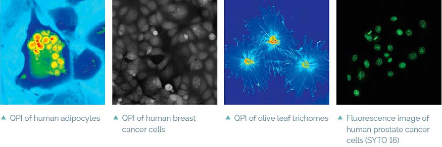
  • Label-free – no staining is needed, simple sample preparation, observation
  • of live cells in their native environment, no photobleaching problems
  • Low phototoxicity – low light power density (107× lower than fluorescence
  • microscopy) allows long-term observations (for days)
  • Coherence-gating effect – Q-PHASE special feature enables it to observe samples
  • even in scattering media (phospholipid emulsions, extracellular matrices, etc.)
  • Multimodality – fully integrated fluorescence module, simulated DIC and
  • brightfield which enables automatic multimodal imaging of the sample
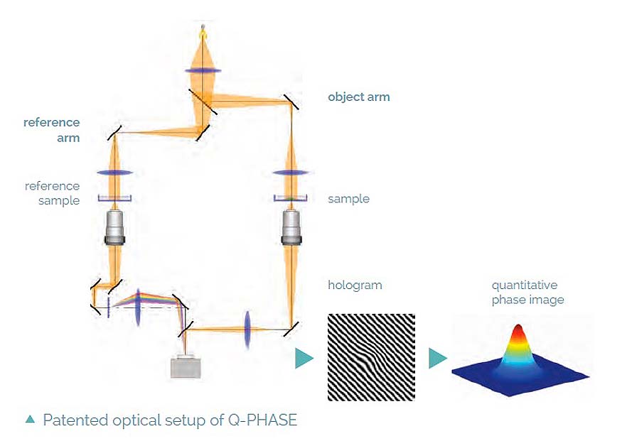
  • High-quality QPI – unique Q-PHASE’s optical setup allows using incoherent illumination
  • which provides extraordinary imaging quality without any compromises
  • Lateral resolution of conventional microscopes (up to 2× better when compared to common laser-based approaches or pinhole spatial filtering based techniques)
  • Fast acquisition – the use of off-axis holographic approach makes Q-PHASE a single-shot instrument, thus enabling imaging of very fast cell dynamics
  • Full motorization – focusing, sample stage, objective exchange, fluorescence
  • filters
  • Automated multidimensional acquisition – time-lapse, channel, position, Z-stack
  • Simple image segmentation and processing – comparable to fluorescence data processing
  • Quantitative – phase values can be recalculated (e.g. to cell dry-mass density (pg/μm2) or direct topography) with nanometer sensitivity (usually non-biological samples with homogeneous refractive index distribution)
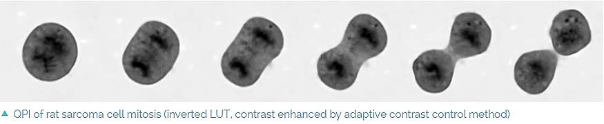
  • High phase detection sensitivity – is able to detect even the smallest changes in axial direction, very sensitive detection of morphology or position changes

Fluorescence module

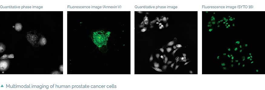
The Q-PHASE can combine holographic microscopy with fluorescence microscopy. This powerful combination provides the possibility to verify structures or processes observed in QPI with fluorescence microscopy in the same field of view using a single instrument. For example, morphological and position changes prior to cell death can be observed in QPI followed by fluorescence verification of cell death types (see images below) [4]. This approach greatly reduces the phototoxicity and photobleaching problems of fluorescence imaging and it allows long-term observations.

The focus plane in both methods is located at the same position. This allows easy and fast switching between the two imaging methods at the same conditions and time points. Multiple fluorescence channels are possible with motorized channel exchange for automated multidimensional measurements.

The illumination can be implemented by using liquid light guide coupled solid state light sources or a xenon arc lamp. Multidimensional image acquisition combining holography and fluorescence is fully integrated in the Q-PHASE’s software. A fluorescence module is attached to the side port of the Q-PHASE, which can alternatively be used for other imaging techniques.

6864

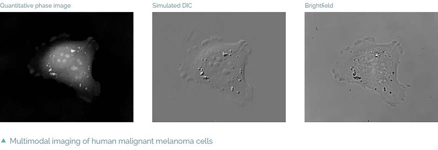
Intrinsic imaging modes

Complementary image contrast can be obtained simply by numerical processing of the acquired phase images. In this way simulated DIC images can be produced with adjustable shear and displayed in real time. Another possibility is brightfield imaging which can be simply achieved by closing the reference arm of the microscope. In summary, the Q-PHASE offers multiple imaging modes widely used in biological research such as fluorescence or DIC integrated in a single instrument and supported by the Q-PHASE’s software allowing fully automated multimodal imaging.

6865

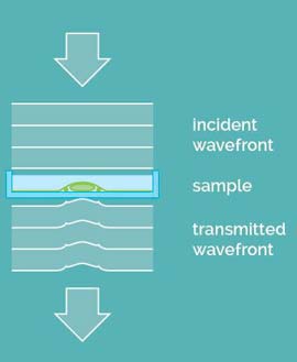
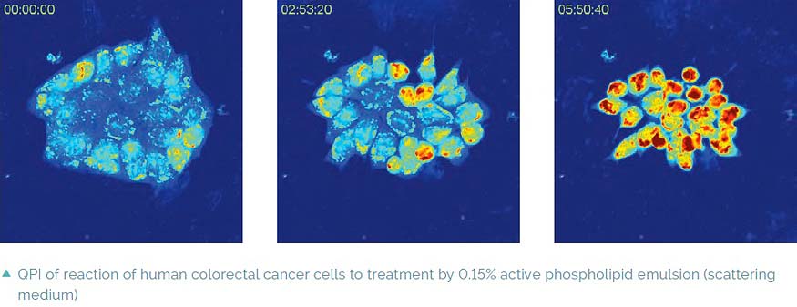
Imaging in scattering media

A special feature of Q-PHASE is coherence-gating, a well-known effect in optical coherence tomography which enables observations of samples even in scattering media. This effect is induced by using incoherent light in the unique patented setup of Q-PHASE. Its transmitted-light configuration enables it to effectively suppress the light which was scattered by the environment in defocused planes and to only use unscattered light for imaging. In this way, cells can be observed even in moderately scattering non-transparent substances such as an active phospholipid emulsion.

6866

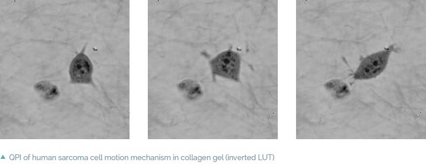
Imaging in extracellular matrices

The coherence-gating effect can also be beneficial when imaging cells in extracellular matrices such as collagen gel. Extracellular matrices mimic an in vivo environment making the study of the cell’s dynamic reactions to its surroundings more realistic. Usually it is used as a biological test for cancer cell invasivity and ability to metastasize. The Q-PHASE microscope enables one to record a mechanism of cell motion and interactions between extracellular matrix fibers and cells with high contrast and without any additional staining.

6867

Applications

Applications

Many different types of samples can be observed including adherent cells in monolayers, thin tissue sections or plant samples. The sample preparation is quite straightforward; simplypour the cell suspension into the observing chamber with no further procedures needed. The cells can also be seeded into the perfusion chamber and perfusion system with the ability to apply different treatments. Cell reactions to the treatment can then be observed online.

Application examples

  • Cell life cycle, cell proliferation, cell differentiation, cell viability, counting
  • Cell dry mass evaluation, cell growth, changes in cell dry mass distributions
  • Cell morphology changes, intracellular processes
  • Cell motility, tracking
  • Cell interactions, cell co-cultures
  • Fast cellular processes
  • Testing reactions of cells to a specific treatment, cytotoxicity
  • Imaging in scattering non-transparent media (e.g. phospholipid emulsions), imaging in 3D environments (e.g. collagen extracellular matrix)
  • Multimodal imaging (QPI automatically correlated with fluorescence,DIC or brightfield)

  6862

6863

Technical Specification

Technical Specification

Microscope configuration transmission inverted microscope
Microscopy techniques holography (quantitative phase imaging), epifluorescence, simulated DIC, brightfield
Objectives magnification 4× to 60×
Objective turret 6-position, motorized exchange
Light source halogen lamp
Operating wavelength 650 nm
Sample stage motorized, 130 mm × 90 mm travel range
Focusing motorized objective turret, 8 mm travel range
Piezo-focusing optional, multiple travel ranges available
Lateral resolution 3.3 μm with 4× NA 0.1 objective 0.57 μm with 60× NA 1.4 objective
Field of view objective dependent, up to 950 μm × 950 μm with 4× objective
Acquisition framerate 5.5 fps at full frame (option: higher framerates possible)
Reconstructed phase image size 600 px × 600 px
Illumination power at sample plane down to 0.2 μW/cm2
Phase detection sensitivity down to 0.0035 rad (0.7 nm at Δn = 0.5) Δn - difference between refractive indexes of sample and surrounding media
Power 230 V/50 Hz (120 V/60 Hz optional), 2300 VA
Dimensions (W × L × H) 1100 mm × 950 mm × 1620 mm microscope with incubator 2515 mm × 974 mm × 1620 mm total with operator table
Weight 350 kg (including microscope table, epi-fluorescence attachment and microscope incubator)


Field and aperture diaphragms
Side port available for fluorescence module or other additional techniques
Microscope table with anti-vibration suspension
Control panel with multifunctional touchscreen, sample stage joystick and rotary knobs
Microscope incubator with computer temperature setting and temperature data logging
Incubation chamber for precise and long-term control of temperature, humidity and CO2 concentrations.


References

References

[1] US patent No. 8526003. [2] T. Slabý et al.: Off-axis setup taking full advantage of incoherent illumination in coherence-controlled holographic microscope. Optics Express 21, 2013, 14747.

[3] H. Janeckova et al.: Proving tumour cells by acute nutritional/energy deprivation as a survival threat: a task for microscopy. Anticancer Research 29, 2009, 2339–2345.

[4] J. Balvan et al.: Multimodal holographic microscopy: distinction between apoptosis and oncosis. PLOS ONE, 2015 (submitted).

[5] R. Barer: Interference microscopy and mass determination. Nature 169, 1952, 366–367.

[6] H. Davies, M. Wilkins: Interference microscopy and mass determination. Nature 169, 1952, 541.