Type
Sandwich ELISA, Biotin-labelled antibody
Applications
Serum, Plasma-Citrate, Cell culture supernatant
Sample Requirements
50 µl/ well (1:10 prediluted)
Shipping
At ambient temperature. Upon receipt, store the product at the temperature recommended below.
Storage/Expiration
Store the complete kit at 2–8°C. Under these conditions, the kit is stable until the expiration date (see label on the box).
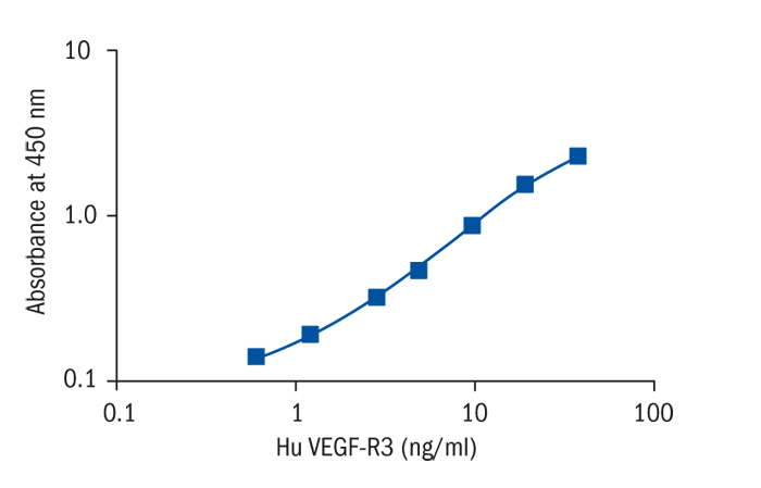
Calibration Curve
Calibration Range
0.6–40.0 ng/ml
Limit of Detection
0.03 ng/ml
Intra-assay (Within-Run)
CV = 5.7%
Inter-assay (Run-to-Run)
CV = 5.0%
Research topic
Cytokines and chemokines and related molecules
Summary
The main function of the lymphatic system is to return plasma back to the circulating blood. Plasma fluid, macromolecules and cells, such as leucocytes and activated antigen-presenting cells, enter the blind-ended lymphatic vessels. Lymph is then transported toward collecting lymphatic vessels and is returned to the blood circulation.
First growth factors and molecular markers specific for lymphatic vessels were discovered only about ten years ago. This is rather astonishing, given the importance of the lymphatic system in maintaining fluid homeostasis and its involvement in the pathogenesis of many diseases, including cancer. Scientific studies over the past years have revealed a signal-transduction system for lymphatic endothelial cell growth, migration and survival. This system is formed by VEGF-C and VEGF-D and their shared receptor VEGFR-3/FLT-4. Homozygous deletion of VEGFR-3 in mice leads to defects in blood-vessel remodelling and embryonic death at mid-gestation, indicating an early vascular function for the receptor. Furthermore mouse studies suggest that upregulation and activation of VEGFR-3 have functional roles in sprouting angiogenesis and that VEGFR-3 is an important effector of the vascular phenotype resulting from Notch inhibition.
Heterozygous missense point mutations leading to receptor kinase inactivation have been found in VEGFR-3 of patients with Milroy disease, a rare autosomal dominant lymph oedema. Metastatic tumour spread through the blood or lymphatic vessels in human cancer, often associated with regional lymph-node metastasis, represents the most important prognostic factor for carcinoma patients. Several studies have shown positive correlations between VEGF-C, VEGF-D and VEGFR-3 expression and vascular invasion, lymphatic vessel and lymph node involvement. VEGF-C expression in tumour cells may be induced by growth factors or proinflammatory cytokines. High levels of VEGF-C or VEGF-D also enhance lymphatic metastasis in various experimental models. In some tumours, processed VEGF-C or VEGF-D may be generated, which mainly target VEGFR-3, but also VEGFR-2, which is often upregulated in tumour blood vessels.
In lymphoma patients, high expression of VEGFR-3 can been found in lymph endothelial cells isolated from the tumour tissue. In corneal inflammation mouse models, dendritic cells expressing both VEGFR-3 and VEGF-C could be detected, suggesting that immune cells may respond to lymphangiogenic signals and induce lymphangiogenesis.
Lymphangiogenesis research has revealed important therapeutic options for human diseases such as lymph oedema and other oedemas that will enter clinical development in the near future. The importance of lymph-node metastasis in the spread of cancer to distant organs needs better understanding before applying the newly acquired knowledge to patients. In this context, the roles of VEGF-C and VEGFR-3 upregulation in tumour angiogenesis need to be explored for additional therapeutical application.
The sandwich ELISA for detecting and reliable measuring of solubilized VEGFR-3 from tumour tissue, lymphatic vessels and different kind of endothelial cells will certainly help to understand the mechanisms of lymphatic metastasis including the identification of tumour determinants that are important for the spread of tumour cells.
Find documents for the lot
Example Instructions for Use (RUO)
Example Instructions for Use (RUO)
Safety Information (RUO)
MSDS (RUO)